
Reasons for Knee Revisions
What to do if your knee replacement is not right
Most knee replacement patients do really well, but a small number of knee replacements can have problems with pain, stiffness, swelling, and/or instability. We will discuss:
Mechanical Loosening
Infection
Stiffness
Instability
Fracture
Wrong diagnosis

Mechanical Loosening
Mechanical loosening typically causes start-up pain from a loose implant shifting or settling down when a patient first puts weight on their knee joint. Start-up pain is where a patient has been walking a fair bit early in the day, then sits down for a short time, and then stands up and has pain with weight-bearing on that knee. The patient cannot trust that their knee replacement will hold them up, and they have to gently stand there for a few seconds to allow their knee to adjust before they can safely walk. Mechanical loosening can occur with both cemented and press-fit implants. Possible tests to diagnose mechanical loosening include radiolucent lines around the implants on x-rays, changes in implant positioning on x-rays, inappropriate bone formation around the implants on x-rays, stress reactions on metal-suppressed MRI, and increased uptake on a nuclear bone scan.
A stress fracture around a knee replacement can present similarly to mechanical loosening with start-up pain but with no radiographic signs of implant loosening. Patients may have a preceding episode of an unusual amount of activity.

Infection
Infection can be obvious or subtle. When a knee replacement is infected with purulent bacteria (like staph), the knee becomes swollen, painful, and warm. The knee may start to drain fluid. The diagnosis is pretty easy to make.
When a knee replacement is infected with an indolent bacteria (slow-growing), the knee may not have any symptoms or just mild symptoms like occasional vague pain.
The workup for a possibly infected knee replacement is mostly obtaining some fluid from inside the knee joint (aspiration) and sending that fluid for analysis of C-reactive protein, cell count, alpha-defensin, and culture. Not all infected knees grow bacteria in their cultures.
Infected knee replacements hurt because of the swelling and implant loosening.

Stiffness
The best predictor of post-op range of motion is pre-op range of motion, but some patients do get stiff after their knee replacement. If a patient does not bend their knee past 90 degrees in the first 6 weeks, I usually recommend we do a manipulation. If a patient is ultimately unhappy with their range of motion, I recommend waiting for at least one year after their initial surgery and then reoperating. During that second surgery, I remove the scar tissue and free up the knee motion.

Knee Instability
Knee instability occurs when the femoral component and tibial component do not move as intended, as in paradoxical rollback and condylar lift-off. Patients may have good range of motion when their leg is dangling over the edge of a table but have difficulty with squats and raising from a chair.
Knee instability usually requires removing the knee implants and implanting new implants with more constraint.
Fracture
Trauma, such as falling down, can break the bone around the knee replacement and require more surgery. Knee replacements are not fragile, but some patients are fragile in their 7th and 8th decades of life.
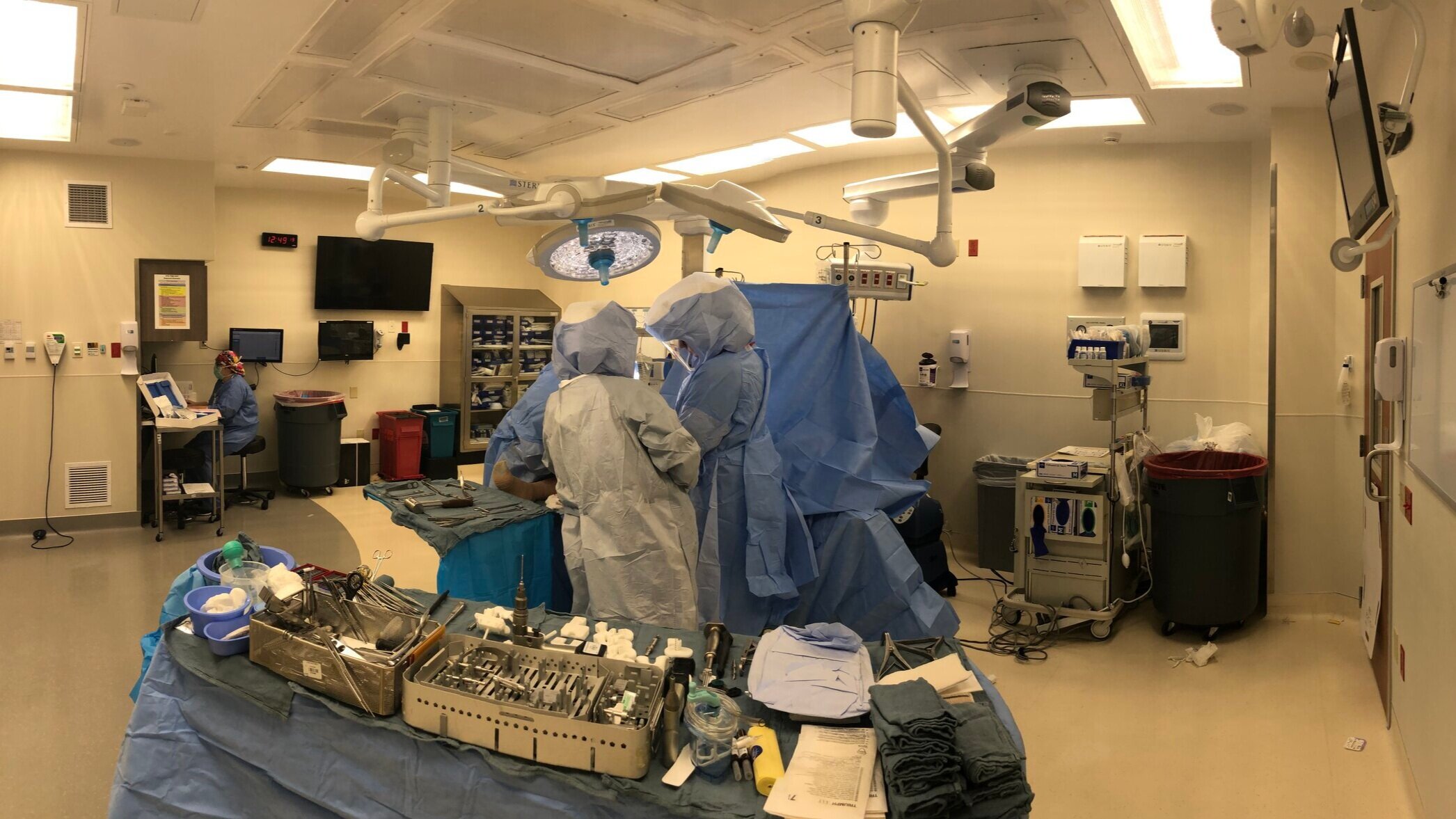
Fractures typically require more surgery to either repair the fracture or revise the implant.
Wrong initial diagnosis
When a patient has the same pain before and after their knee surgery, then it is possible that their pain is coming from something other than their knee. Occasionally, lower back problems and hip arthritis can be perceived as mostly knee pain. If a patient with hip arthritis complains of knee pain and x-rays show knee arthritis, some patients may get a knee replacement instead of the hip replacement that they really needed.
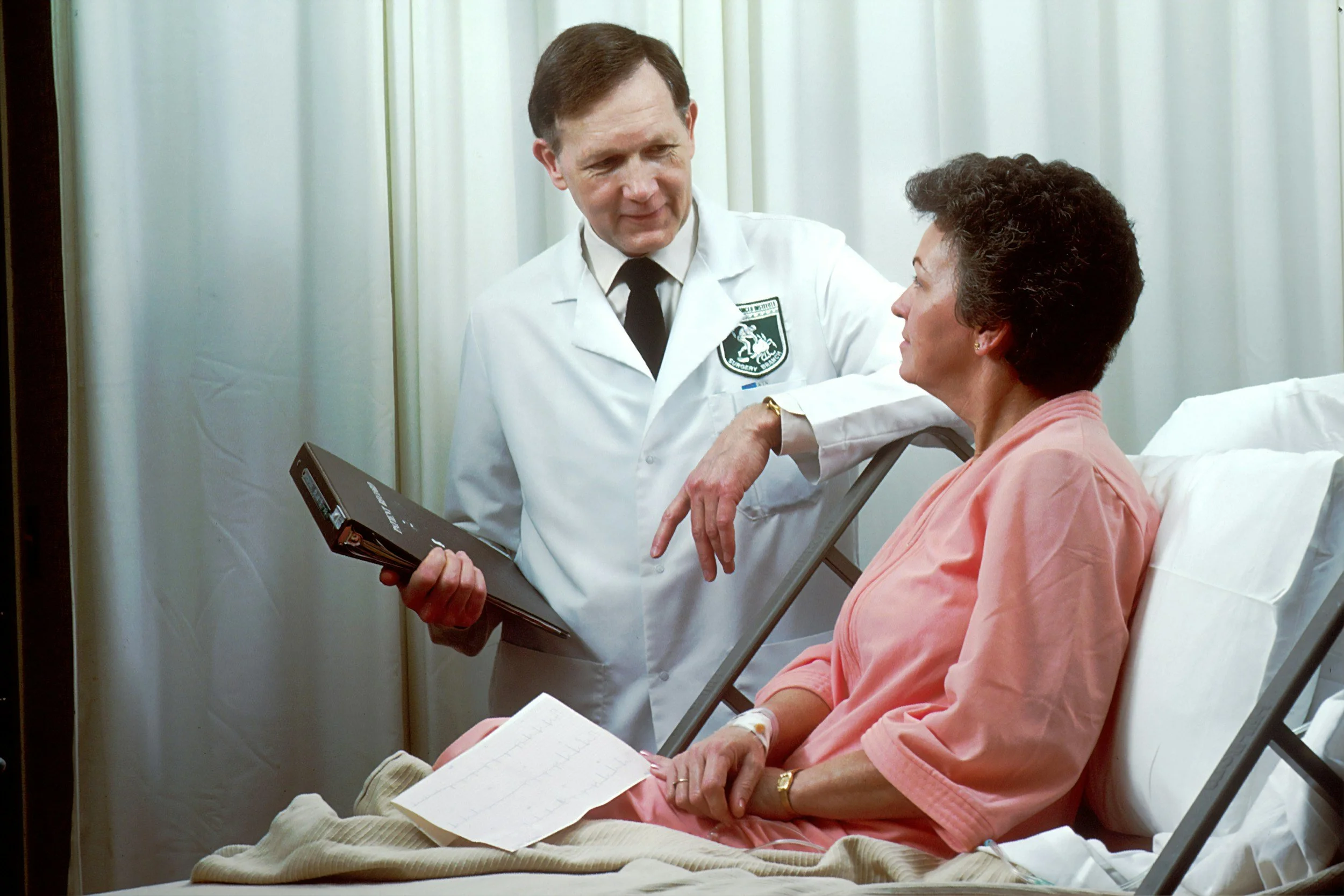
When a knee replacement still hurts, it is important for the surgeon to go back to the beginning of the knee problem and think outside the box.

Work up
The workup of a painful knee replacement includes x-rays of the knee, hip, and L spine; aspiration of the knee; metal-suppressed MRI of the knee; and/or bone scan.